


PNC-27: Structural Characterization, HDM-2-Dependent Membrane Targeting, and Tumor Cell Necrosis Research
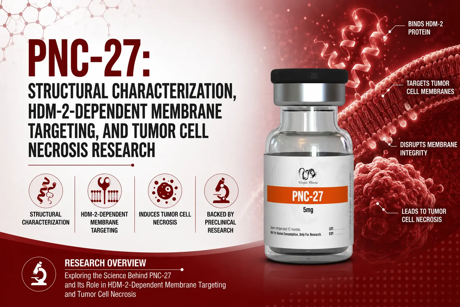
PNC-27 is a synthetic chimeric peptide engineered to engage the HDM-2 protein (the human homolog of murine MDM-2) expressed on the plasma membranes of cancer cells. It combines an HDM-2-binding sequence from the p53 tumor suppressor protein (residues 12–26) with a cell-penetrating leader sequence derived from the Drosophila Antennapedia homeodomain protein. This article covers its structure, proposed membrane-pore mechanism, and preclinical research findings, all of which remain at the in vitro, ex vivo, and animal-model stage — no human clinical trial data exists for PNC-27 at this time.
Key Facts at a Glance
| Property | Detail |
|---|---|
| Structure | Chimeric peptide: p53-derived HDM-2-binding domain (residues 12–26) + penetratin cell-penetrating sequence |
| Primary target | Membrane-localized HDM-2 (preferentially expressed on transformed/cancer cells) |
| Proposed mechanism | HDM-2-dependent transmembrane pore formation ("poptosis"), distinct from apoptosis/necroptosis |
| p53 status dependency | Reported to act independently of intracellular p53 status in tested cell lines |
| Related compound | PNC-28 (same HDM-2-binding domain, shorter penetratin leader) |
| Evidence type | In vitro (human cancer cell lines), one ex vivo primary human tumor study, one cited in vivo mouse xenograft finding. No human clinical trials. |
Table of Contents
- What Are Copper Peptides?
- Historical Development
- Coordination Chemistry and Mechanisms
- A Note on Evidence Quality
- GHK-Cu: Extracellular Matrix and Collagen Research
- GHK-Cu: Wound Repair Across Preclinical and Clinical Models
- GHK-Cu: Antioxidant and Anti-Inflammatory Signaling
- GHK-Cu: Neuromodulatory and Cognitive Research
- AHK-Cu: Dermal Fibroblast and Hair Follicle Research
- DAHK-Cu: Redox Biology and Copper Transport
- Comparing the Three Copper Peptides
- References
Historical Development
PNC-27's conceptual foundation traces to early research identifying that peptides modeled on the amino-terminal MDM-2-binding domain of p53 showed selective cytotoxicity toward transformed cells in vitro, without harming normal cell populations.
These foundational in vitro observations established the rationale for targeting membrane-associated HDM-2 as a cancer-selective approach [1]. PNC-27 was originally conceived as a nuclear decoy peptide intended to competitively block the p53-HDM-2 interaction inside the nucleus. However, experimental observations revealed its primary cytotoxic activity occurs at the plasma membrane rather than intranuclearly, leading researchers to propose the membrane-targeted, HDM-2-colocalization mechanism now supported by multiple independent structural and imaging studies [3][9].
Proposed Mechanism: "Poptosis"
PNC-27 is proposed to kill cancer cells through a two-step, HDM-2-dependent process distinct from classical apoptosis: formation of 1:1 PNC-27/HDM-2 complexes at the membrane, followed by temperature-dependent dimerization into transmembrane pore structures.
The resulting pores are proposed to enable rapid, explosive release of intracellular contents — a process termed "poptosis" to distinguish it from apoptotic and necroptotic pathways. Research suggests poptosis may operate independently of caspase activation and intracellular p53 signaling, since tumor cell lines lacking p53 expression have remained susceptible to PNC-27-induced necrosis in tested models [5].
Research Findings by Study
HDM-2 Binding Conformation and Antibody-Blocking Validation
Computational modeling suggested the p53-derived segment of PNC-27 adopts a three-dimensional configuration matching p53 residues in HDM-2-bound crystal structures, supporting a p53-mimicry binding hypothesis. In cultured human cancer cells (in vitro), incubation with a monoclonal antibody targeting the p53-binding site of HDM-2 substantially blocked PNC-27-induced necrosis, while control immune sera did not — supporting the model that PNC-27 engages the amino-terminal p53-binding domain of membrane HDM-2 as a prerequisite for pore formation [3].
Intact Peptide, Not Fragments, Drives Membranolysis
Using dual-terminal fluorescent labeling (FITC/TRITC) to track PNC-27 during membrane interactions with MCF-7 human breast cancer cells and MCF-10-2A untransformed human breast cells (both in vitro), researchers found the intact, full-length peptide — not proteolytic fragments — was present at the membrane during lysis events in cancer cells. Untransformed cells showed peptide degradation without lysis [4].
Membrane HDM-2 Expression Is the Selectivity Determinant
Researchers artificially introduced membrane-associated HDM-2 expression into normal, untransformed human cells that do not naturally express it. These transfected cells became susceptible to PNC-27-induced lysis, while untransfected controls remained viable — supporting the hypothesis that membrane HDM-2 presence, not general cell phenotype differences, determines PNC-27's cancer selectivity [2].
Leukemia Cell Line Activity (p53-Independent)
Using the K562 human chronic myelogenous leukemia cell line (in vitro, notably lacking p53 expression) with murine leukocytes as normal controls, researchers detected membrane HDM-2 on the leukemia cells via flow cytometry and immunohistochemistry. PNC-27 exposure produced tumor cell necrosis consistent with pore formation, while control murine leukocytes did not lyse — supporting a p53-independent mechanism driven by membrane HDM-2 presence rather than intracellular p53 status [5].
Acute Myelogenous Leukemia: Three Cell Line Study
Across three human AML cell lines (U937, OCI-AML3, HL-60; all in vitro), all three showed elevated membrane HDM-2 by flow cytometry, and PNC-27 exposure produced measurable LDH release within 4 hours, consistent with rapid membrane pore formation. Annexin V and caspase-3 marker patterns were interpreted as consistent with necrosis rather than apoptosis. Normal hematopoietic cells tested in parallel did not show comparable cytotoxicity [7].
PNC-28 and the Role of the Penetratin Domain
PNC-28, sharing PNC-27's HDM-2-binding domain but with a shorter penetratin leader, was studied in human pancreatic cancer cells (in vitro) to isolate the penetratin sequence's contribution. Findings suggested the penetratin component may direct cell death toward necrosis rather than apoptosis — a distinction potentially relevant to lysis efficiency across this peptide class [6].
Ex Vivo Primary Human Ovarian Tumor Tissue
This study used ex vivo primary human tumor specimens rather than an established cell line — a meaningfully stronger evidence context than immortalized cell line work, though still preclinical.
PNC-27 retained selective cytotoxic activity against primary ovarian tumor cells derived directly from patient specimens, while normal human ovarian epithelial cells remained unaffected under comparable ex vivo conditions [10].
Mitochondrial Targeting: A Secondary Mechanism
Using MIA-PaCa-2 human pancreatic carcinoma cells (in vitro) and immunoelectron microscopy with gold-particle-conjugated anti-PNC-27 antibodies, researchers found the peptide detectable on mitochondrial membranes following plasma membrane pore formation, alongside mitotracker dye loss (consistent with mitochondrial disruption) and retained lysotracker signal. Normal human fibroblast controls did not show comparable mitochondrial disruption — suggesting a dual plasma-membrane-plus-mitochondrial mechanism [9].
In Vivo Mouse Xenograft Findings (Cited in Review)
A 2024 mechanistic review synthesizing the poptosis literature cited findings from nude mouse xenograft models in which PNC-27 was reported to reduce growth of highly metastatic pancreatic tumors and stem-cell-enriched AML tumors transplanted into bone marrow, with no detectable off-target toxicity in normal tissue in those models [8]. This represents the only in vivo (live animal) evidence referenced across the available literature for this compound — all other findings summarized here are in vitro or ex vivo.
Evidence Summary: Study Type and Model by Research Area
| Research Finding | Model | Evidence Tier |
|---|---|---|
| Selective cytotoxicity to transformed cells | Human cancer cells, cultured | In vitro |
| HDM-2 antibody-blocking of necrosis | Cultured human cancer cells | In vitro |
| Intact-peptide membranolysis | MCF-7 / MCF-10-2A human cell lines | In vitro |
| HDM-2 transfection selectivity test | Human cell lines | In vitro |
| p53-independent leukemia necrosis | K562 human cell line; murine leukocyte controls | In vitro |
| AML cell line necrosis (3 lines) | U937, OCI-AML3, HL-60 human cell lines | In vitro |
| Penetratin domain and cell death pathway | Human pancreatic cancer cells (PNC-28) | In vitro |
| Primary ovarian tumor cytotoxicity | Patient-derived primary tumor tissue | Ex vivo (human tissue) |
| Mitochondrial disruption mechanism | MIA-PaCa-2 human cell line | In vitro |
| Tumor growth reduction, off-target toxicity | Nude mouse xenografts | Animal (in vivo) |
Storage and Stability
| Condition | Recommendation |
|---|---|
| Lyophilized form | Store frozen (-20°C) for long-term stability |
| Reconstituted solution | Refrigerate (2–8°C); use within the research protocol's defined window |
| Light exposure | Store protected from light |
| Handling | Avoid repeated freeze-thaw cycles to preserve peptide integrity |
Frequently Asked Questions
What is PNC-27?
PNC-27 is a synthetic chimeric peptide combining a p53-derived HDM-2-binding domain with a cell-penetrating penetratin sequence, studied for selective cytotoxicity toward cancer cells that express HDM-2 on their plasma membranes.
How does PNC-27 kill cancer cells?
Research proposes a two-step mechanism termed "poptosis": PNC-27 forms 1:1 complexes with membrane-bound HDM-2, which then dimerize into transmembrane pores, leading to rapid loss of intracellular contents distinct from classical apoptosis.
Does PNC-27 require functional p53 to work?
Research indicates PNC-27 can induce necrosis in cell lines lacking p53 expression, suggesting its mechanism depends primarily on membrane HDM-2 presence rather than intracellular p53 status.
Has PNC-27 been tested in humans?
No human clinical trial data currently exists for PNC-27. Available evidence comes from in vitro cell line studies, one ex vivo primary human tumor tissue study, and cited findings from mouse xenograft models.
What is the difference between PNC-27 and PNC-28?
Both share the same HDM-2-binding domain derived from p53, but PNC-28 incorporates a shorter penetratin leader sequence. Research suggests the penetratin domain influences whether cell death proceeds via necrosis or apoptosis.
Why doesn't PNC-27 affect normal cells?
Research indicates normal, untransformed cells generally lack membrane-localized HDM-2, which appears to be the critical determinant of PNC-27's selectivity; artificially introducing membrane HDM-2 into normal cells made them susceptible to PNC-27-induced lysis.
Has PNC-27 been studied in blood cancers?
Yes. In vitro studies in leukemia cell lines, including K562 chronic myelogenous leukemia and three acute myelogenous leukemia cell lines, found membrane HDM-2 expression and PNC-27-induced necrosis, with normal blood cell controls unaffected.
Does PNC-27 affect mitochondria?
An in vitro study found evidence of PNC-27 entering cells after plasma membrane pore formation and disrupting mitochondrial membranes, suggesting a secondary mechanism alongside its primary plasma-membrane activity.
What is HDM-2?
HDM-2 is the human homolog of murine MDM-2, a protein that normally regulates the tumor suppressor p53; research suggests it is also expressed on the plasma membrane of many cancer cell types, distinct from its typical intracellular role.
Has PNC-27 shown activity in primary tumor tissue, not just cell lines?
Yes, in one ex vivo study using patient-derived primary ovarian tumor specimens, where PNC-27 retained selective cytotoxic activity while sparing normal ovarian epithelial cells under the same conditions.
Key Takeaways
- PNC-27 targets HDM-2 specifically when it is expressed on the plasma membrane of cancer cells — a target largely absent from normal cell membranes in tested models.
- Its proposed mechanism, "poptosis," involves HDM-2-dependent transmembrane pore formation, mechanistically distinct from apoptosis, and does not appear to require functional intracellular p53.
- Research spans solid tumor cell lines, multiple leukemia cell lines, one ex vivo primary human ovarian tumor study, and secondary mitochondrial disruption findings.
- All available evidence is preclinical: in vitro cell line and ex vivo tissue studies, plus one cited in vivo mouse xenograft finding. No human clinical trial data currently exists for PNC-27.
References
- Kanovsky M, Raffo A, Drew L, et al. Peptides from the amino terminal mdm-2 binding domain of p53, designed from conformational analysis, are selectively cytotoxic to transformed cells. Proc Natl Acad Sci USA. 2001;98(22):12438-43. https://pubmed.ncbi.nlm.nih.gov/11606716/
- Sarafraz-Yazdi E, Mumin S, Cheung D, et al. PNC-27, a Chimeric p53-Penetratin Peptide Binds to HDM-2 in a p53 Peptide-like Structure, Induces Selective Membrane-Pore Formation and Leads to Cancer Cell Lysis. Biomedicines. 2022;10(5):945. https://doi.org/10.3390/biomedicines10050945
- Sarafraz-Yazdi E, Bowne WB, Adler V, et al. Anticancer peptide PNC-27 adopts an HDM-2-binding conformation and kills cancer cells by binding to HDM-2 in their membranes. Proc Natl Acad Sci USA. 2010;107(4):1526-31. https://pubmed.ncbi.nlm.nih.gov/20080680/
- Sookraj KA, Bowne WB, Adler V, et al. The anti-cancer peptide, PNC-27, induces tumor cell lysis as the intact peptide. Cancer Chemother Pharmacol. 2010;66(2):325-31. https://pubmed.ncbi.nlm.nih.gov/20182728/
- Davitt K, Babcock BD, Fenelus M, et al. The anti-cancer peptide, PNC-27, induces tumor cell necrosis of a poorly differentiated non-solid tissue human leukemia cell line that depends on expression of HDM-2 in the plasma membrane of these cells. Ann Clin Lab Sci. 2014;44(3):241-8. https://pubmed.ncbi.nlm.nih.gov/25117093/
- Bowne WB, Sookraj KA, Adler V, et al. The penetratin sequence in the anti-cancer PNC-28 peptide causes tumor cell necrosis rather than apoptosis of human pancreatic cancer cells. Ann Surg Oncol. 2008;15(12):3588-3600. https://pubmed.ncbi.nlm.nih.gov/18931881/
- Thadi A, Morano WF, Khalili M, et al. Targeting Membrane HDM-2 by PNC-27 Induces Necrosis in Leukemia Cells But Not in Normal Hematopoietic Cells. Anticancer Res. 2020;40(9):4857-4867. https://www.researchgate.net/publication/344117616
- Pincus MR, Silberstein M, Zohar N, Sarafraz-Yazdi E, Bowne WB. Poptosis or Peptide-Induced Transmembrane Pore Formation: A Novel Way to Kill Cancer Cells without Affecting Normal Cells. Biomedicines. 2024;12(6):1144. https://www.ncbi.nlm.nih.gov/pmc/articles/PMC11201261/
- Krzesaj P, Adler V, Feinman RD, et al. Anti-Cancer Peptide PNC-27 Kills Cancer Cells by Unique Interactions with Plasma Membrane-Bound hdm-2 and with Mitochondrial Membranes Causing Mitochondrial Disruption. Ann Clin Lab Sci. 2024;54(2):137-148. https://pubmed.ncbi.nlm.nih.gov/38802154/
- Orynbayeva Z, Senkalı D, Bhatt DL, et al. Ex vivo Efficacy of Anti-Cancer Drug PNC-27 in the Treatment of Patient-Derived Epithelial Ovarian Cancer. Ann Clin Lab Sci. 2015;45(6):650-656. https://www.annclinlabsci.org/content/45/6/650.long
Articles
What is Post Cycle Therapy and how to do it after each steroid cycle.

This article organizes the available research by mechanism and study type, distinguishing behavioral findings from molecular and vascular research.
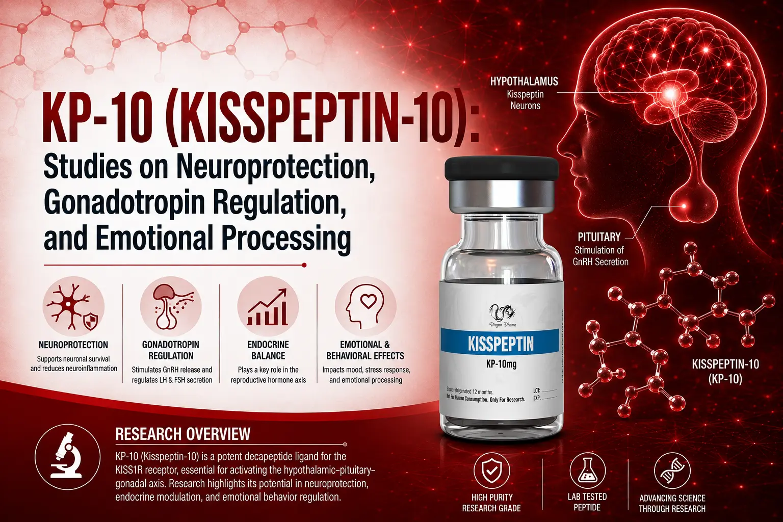
This article covers its role as an endogenous GPR54 (KISS1R) ligand regulating the hypothalamic-pituitary-gonadal axis, its emerging neuroprotective research, and its studied effects on appetite and emotional/sexual brain processing—including a correction where human clinical data was found labeled as "murine models" in the source material, alongside a species contradiction (mice don't have a menstrual cycle) that revealed the mislabeling.
Customer Reviews
Please leave your review on products or service below.
Thank you beforehand.